What can be measured in my bone biopsy
This is the slide from my iliac crest biopsy, stained with haematoxylin and eosin: the same tissue the molecular panel came from. Every number on this page is measured on the image itself, calibrated against its own scale bar. Below is what can be counted and measured in it and, in the same detail, where what you can claim by looking at it runs out. Nothing here is a diagnosis.
Field of 831 by 296 microns. 767 nuclei were segmented with a trained model, with a median diameter of 7.2 microns. The tables on this page are the full text alternative to the image.
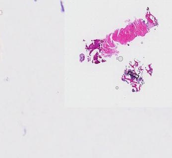
El recuadro que marca el campo es un trazo de 1 píxel sobre un lado corto de 5 a 6 píxeles. Medirlo ahí arrastra un error del 20% en el lado corto, y la escala que sale va de 35 a 58 µm/px.
Findings
| Field | 831 × 296 µm · 0.454 µm/px ± 1% |
| Tissue | 0.183 mm² (0.165–0.221) |
| Trabecular bone | 12–18 % of tissue · 0.023–0.035 mm² |
| Nuclei | 767 · median 7.2 µm (p10–p90 5.1–9.7) |
| Nuclear density | 6721/mm² of cellular mass |
| Effective resolution | 2.3 µm · floor 1.5 µm |
| Bone-tumour interface | processing retraction |
Not assessable in this field
| Neuroendocrine phenotype | chromatin texture below the limit (2.3 µm) |
| Tumour extent in the block | single section |
| Lytic or blastic pattern | one trabecula |
| Grade and proliferation index | whole slide and its own stain |
| Osteocyte density | ≤7 identifiable lacunae (ref. 150–210/mm²) |
| Core length | no scale bar on the overview · report: 6 cores, 4–11 mm |
Ruifrok-Johnston stain deconvolution, Macenko background, nuclei with InstanSeg. The figures have been corrected twice since publication; the history is public in the repository.
What the molecular panel of the same block said
The same tissue shown above was sequenced with a 324-gene panel. These are the findings as the report states them. What they mean for treatment is decided by the medical team, not by this page.
The two amplicons
These are two events, not seven. The genes in each block sit next to each other on the chromosome and amplify together. They were already present in the 2024 primary biopsy, with fewer copies.
Genomic signatures
| Homologous recombination signature | HRDsig negativa (0,02) |
| Microsatellites | Estables |
| Tumour mutational burden | 4 Muts/Mb |
Sample quality
| Tumour cellularity | 80% / 61% |
| Median coverage | 2.153x |
| Estimated ploidy | 2,38 |
One point variant
MUTYH G382D
Allele frequency 43,2%. The report recommends a blood test to determine whether it is inherited or tumour-derived.
No reportable alteration
AKT1 · BRCA1 · BRCA2 · ERBB2 · ESR1 · PIK3CA · PTEN
No reportable alteration. Reliable for point mutations; the panel does not cover ESR1 fusions, single-copy losses or HER2 by immunohistochemistry.